jetOPTIMUS®
jetOPTIMUS® is an innovative cationic nanotechnology developed to improve DNA transfection efficiency in hard-to-transfect cells.
![]()
![]()
Neural cells are indispensable cell models for cutting edge neurobiology research aiming to dissect molecular mechanism at play in physiological and diseases conditions. From neural stem cell lineage differentiation two major sub-populations can be distinguished: neuronal and glial cells (Bond A., et al 2021). Primary neurons can develop and display properties of mature neurons, as well as spontaneously elaborate neurites if maintained under appropriate culture conditions (Li Z., et al 2011). Primary neurons can thus display characteristics of corresponding cells in situ which makes them an interesting model for neurobiological studies. Glial cells are considered as key players in many neuronal processes providing support and protection (Jäckel S., et al. 2017).
Efficient transfection of primary neuronal cells is essential to perform gene functional studies to address many neurobiological questions regarding brain development, genetics, and disease mechanisms. Chemical-based transfection method is the easiest delivery method to ensure reproducible delivery of DNA and mRNA in primary neuronal cells. To ensure successful transfection, it is critical to consider the sensitivity of primary neuron cells to their microenvironmental change such as air exposure, temperature or pH variations, their limited life span (typically days to weeks) and the fact that they do not actively divide. Here we provide recommendations for efficient transfection of most used primary cell from neuronal lineages, including primary mouse and rat cortex neurons, rat hippocampal neurons, human primary astrocytes and IPSC-derived dopaminergic neurons (DopaNeurons). During transfection protocol optimisation for each neuron cell type, we analyzed delivery efficiency and cell viability using our difficult to transfect specific DNA and mRNA transfection reagents, namely jetOPTIMUS® and jetMESSENGER®. JetOPTIMUS® is cationic polymer-based and ensures high transfection efficiency of DNA in various primary cell types with minimal impact on cellular viability and morphology. JetMESSENGER® is lipid-based and enables improved delivery in slow to non-dividing primary cells using mRNA instead of plasmid DNA.
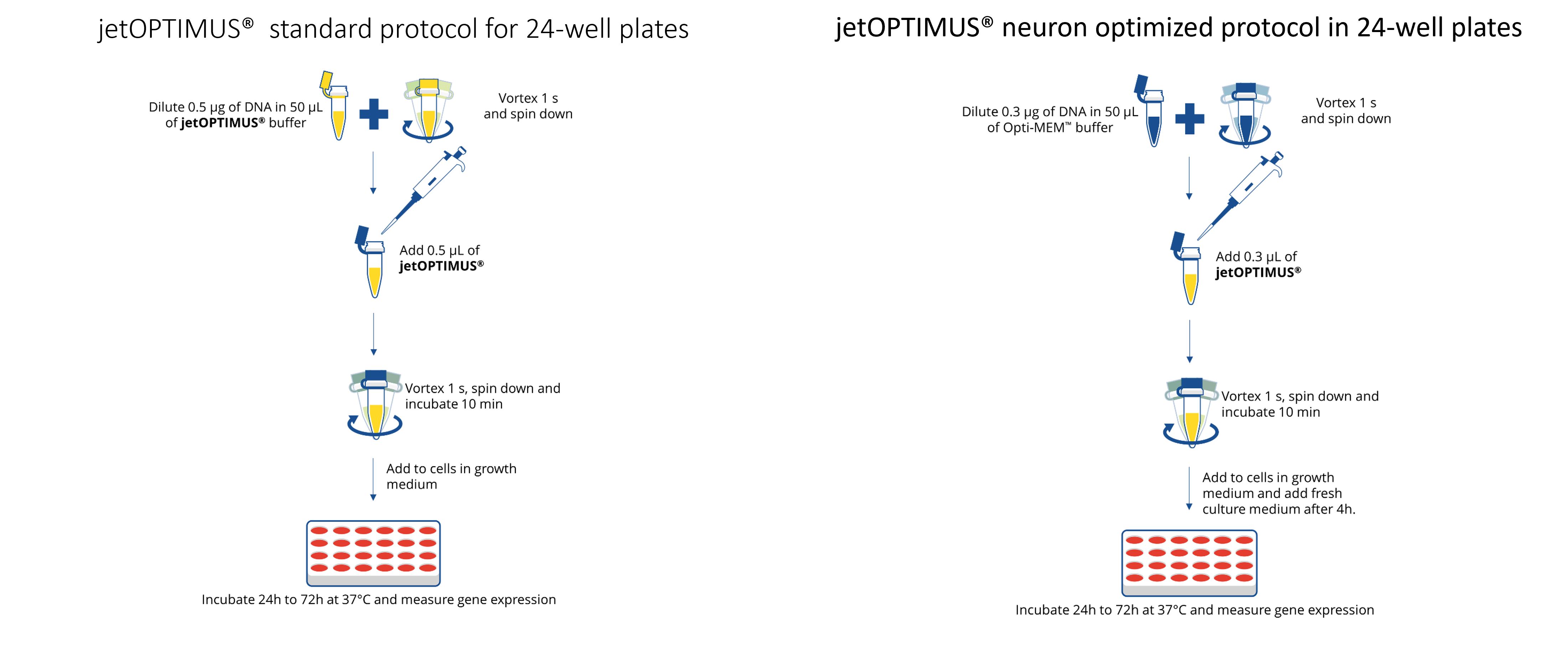
To improve gene expression in differentiated neural cells, such as neurons, we developed cell-specific transfection conditions based on our standard jetOPTIMUS® protocol designed for hard to transfect primary cells and cell lines. In comparison to the standard jetOPTIMUS® protocol, we identified that the DNA amount as well as the DNA-to-reagent ratio were key parameters to achieve fold-increase transfection efficiencies across a panel of diverse primary neuronal and glial cells. As illustrated in Figure 1, we recommend decreasing the amount of DNA down from 0.5 ug to 0.3 ug per well of a 24-well plate, as well as decreasing the amount of reagent to keep a DNA-to reagent-ratio of 1:1.
With the neuron-specific transfection protocol, we showed that jetOPTIMUS® improved transfection efficiency of primary cortical neurons while preserving neuronal morphology and axonal prolongations compared to competitor (Figure 2).

Figure 2. Transfection of primary rat cortical neurons using jetOPTIMUS® vs Lipofectamine® 2000. Transfections of thawed primary rat cortex neurons were performed using a plasmid coding for eGFP protein under the control of the CMV promoter in 48-well plates following manufacturer’s recommendations for Lipofectamine® 2000 and jetOPTIMUS®. Transfections were carried out 4 days after cell seeding, in complete culture medium. Transfection efficiency was assessed by fluorescent microscopy 24h post-transfection.
We also tested jetOPTIMUS® on neuroglial cells, specifically in primary human normal astrocytes obtaining very high transfection rate of about 60% compared to competitor (Figure 3). Our results demonstrate that jetOPTIMUS® optimized protocol is a very suitable transfection reagent to delivery DNA in neuronal and glial primary cultures.

Figure 3. Astrocyte transfection using jetOPTIMUS® vs Lipofectamine® 2000. Transfections of primary human normal astrocytes were performed using a plasmid coding for the eGFP protein under the control of the CMV promoter, in 24-well plates following the manufacturer’s recommendations for jetOPTIMUS® and Lipofectamine® 2000. (A) Microscopy analysis was performed 24h post-transfection. (B) Transfection efficiency was assessed by flow cytometry analysis 24 h post-transfection
To increase transfection efficiency for different kind of applications such as functional and biochemical assays, we transfected eGFP mRNA in different types of neuronal and glial cells. As shown in Figure 4, we obtained high transfection rate above 40% for hippocampal and cortical rat primary neurons. Moreover, high transfection efficiency was achieved for dopaNeurons obtained from iPScells, indicating that jetMESSENGER® is a very powerful tool to study reprogramming and differentiation. For human normal astrocytes, we obtained almost 100% of transfection efficiency (Figure 4). These data demonstrate that jetMESSENGER® mRNA transfection reagent manufactured at Polyplus-transfection® is the most efficient delivery method for neuronal and glial cells.
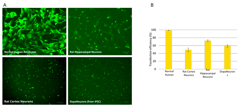
Figure 4. Transfection of different type of neurons using jetMESSENGER®. (A) Representative images of different neuronal and glial cells using jetMESSENGER®. mRNA encoding eGFP (EGFP-mRNA) was transfected with jetMESSENGER® and 4h post-transfection eGFP expression was assessed by fluorescence microscopy. (B) Transfection efficiency was assessed by flow cytometry analysis 24 h post-transfection for astrocytes. For cortex, hippocampal and DopaNeurons, fluorescent microscopy analysis was performed using a fluorescent cell image
Thawed primary rat cortex and hippocampal neurons were respectively purchased from Thermo Fisher Scientific (#A10840-02) and Sigma Aldrich (#R886N-10). Cell culture was performed in Neurobasal medium (#21103-049, Thermo Fisher Scientific) supplemented with B-27 Supplement 50X (1X final, #17504-044, Thermo Fisher Scientific) and GlutaMAX Supplement 200mM (0.5mM final, #35050-038, Thermo Fisher Scientific) following the manufacturers’ recommendations. Poly-D-Lysine solution (#P7886, Sigma) was used for 48-well plate coating before seeding (4.5µg/cm2). Cells were seeded at 100 000 viable cells/cm2 density, with a final volume of 0.5mL of complete culture medium. The culture plates were incubated at 37°C with 5% CO2. 24 hours later, half of the culture medium was removed and replaced with complete culture medium.
iCell® DopaNeurons, human dopaminergic neurons derived from induced pluripotent stem (iPS) were purchased from STEMCELL Technologies (#01279). Cell culture was performed in iCell DopaNeurons Maintenance Medium (100mL, #M1010, STEMCELL Technologies), supplemented with iCell Neural Supplement B (2mL, #M1031, STEMCELL Technologies) and iCell Nervous System Supplement (1mL, #M1031, Fujifilm). Cell culture was prepared following the manufacturers’ recommendations. Poly-L-Ornithin solution (#P4957, SIGMA) and Laminin (#L2020, SIGMA) were used for the vessel coating. For cell seeding, 200 000 viable cells/cm2 were seeded in a 48-well plate, in a final volume of 0.3mL of culture medium. The culture plates were incubated at 37°C with 5% CO2. After 24 hours, 0.3mL of medium was removed and replaced with 0.2mL of fresh medium in each well to obtain a final volume of 0.5mL per well.
Normal human astrocytes (NHA) were purchased from LONZA (#CC-2565). Cell culture was performed in the AGM BulletKit (#CC-3186, LONZA) following the manufacturers’ recommendations. Thawing was carried out directly in a flask and culture vessel was incubated at 37°C with 5% CO2. NHA proliferating culture was assured for experimental use for at least 5 population doublings. During the cell seeding, 10 000 viable cells/well were plated in 24-well plate, in a final volume of 0.5mL of complete culture medium. Plates were incubated at 37°C with 5% CO2 for three days before transfection.
Fresh primary mouse cortex neurons were extracted from C57Bl/6 mice brain. Cell culture was performed in Neurobasal medium (#12348017, Thermo Fisher Scientific) supplemented with 2% of B-27 Supplement (#17504044, Thermo Fisher Scientific), 1% N2 (#17502048, Thermo Fisher Scientific), 1% Penicillin/Streptomycin (#15140122, Thermo Fisher Scientific), 1% L-Glutamine (#25030024, Thermo Fisher Scientific) and 1% fetal bovine serum (Biowest). Poly-D-Lysine solution was used for 48-well plate coating before seeding (4.5µg/cm2). 50 000 viable cells were seeded per well in 24-well plate, in a final volume of 1mL of complete culture medium. The culture plates were incubated 5-7 days at 37°C, with 5% CO2. Cytosine Arabinoside solution (ARA-C) was added once three hours after seeding at 0.8µm final concentration, to stop glial cell proliferation.
Enhanced green fluorescent protein (eGFP) plasmid DNA and mRNA-eGFP were respectively purchased from Polyplus e-Zyvec plasmids (pCMV-eGFP_ez (3.5Kb, #A629.2C) and Tebu-Bio (#L-7201). DNA transfection was carried out with jetOPTIMUS® (#117-01, Polyplus-Transfection®) or Lipofectamine®2000 (#10696343, Thermo Fisher Scientific) and mRNA transfection with jetMESSENGER® (#150-01, Polyplus-Transfection®).
For primary rat cortical neurons, half of the culture medium was removed to transfect in a reduced culture medium volume of 250ul/well. 0.15µg of plasmid was diluted in 25µL of Opti-MEM™ (#31985-062, Thermo Fisher Scientific). DNA solution was vortexed and 0.15µL of jetOPTIMUS® were added. This mixture was vortexed and incubated at room temperature for 10 minutes. 25µL of complexes were added for each well (48-well plate). 4h after transfection, 0.25mL of culture medium were added in each well.
For human normal astrocytes, culture medium was refreshed before transfection. 0.3µg of DNA were diluted in 30µL of Opti-MEM™. 0.3µL of jetOPTIMUS® were added to DNA solution, vortexed and incubated 10 minutes at room temperature. 30µL of the mixture were added per well containing 0.3mL of culture medium. 4 hours later, culture medium was completely removed and refreshed by 0.5mL of fresh culture medium.
For primary mouse cortex neurons, half of the culture medium was removed to transfect in reduced culture medium volume of 0.5mL/well. 0.3µg of DNA were diluted in 50µL of Opti-MEM™. 0.3µL of jetOPTIMUS® were introduced on DNA solution, vortexed and incubated 10 minutes at room temperature. 50µL of DNA/jetOPTIMUS® complexes were added per well containing. 4 hours later, 0.5mL of culture medium were added per well.
For primary rat cortex, hippocampal and dopaminergic neurons, half of the culture medium was replaced by same volume of pre-warmed complete culture medium (final volume of 0.5mL/well). 0.15µg of mRNA-eGFP were diluted in 50µL of mRNA Buffer (jetMESSENGER® buffer, Polyplus-Transfection®). mRNA diluted solution was mixed by pipetting up and down. Then, 0.3µL of jetMESSENGER® were added and solution was homogenized by pipetting up and down.
For human normal astrocytes, 0.4µg of mRNA-eGFP were diluted in 50µL of mRNA buffer. After homogenization by pipetting up and down, 1.2µL of jetMESSENGER® were added to the mRNA solution. After mixing, the mixture was incubated 15 minutes at room temperature. 50µL of mRNA-eGFP/jetMESSENGER® complexes were introduced in a well.
For all the cultures, microscopy analysis was performed 24 hours post-transfection. The ZOE Fluorescent Cell Imager (Bio-rad) was used to take pictures, eGFP+ cells number was assessed by manual counting.
For human normal astrocytes, transfection efficiency was assessed by flow cytometry analysis, using the Guava EasyCyte 5HT (Merck-Millipore) 24 hours post-transfection. The ZOE Fluorescent Cell Imager (Bio-rad) was used to observe and take pictures of eGFP+ cells.
1) Bond A. et al., 2021 Current Topics in Developmental Biology, 67-98, 142.
2) Li Z. et al., Comprehensive Biotechnology, Second Edition, 551-63, 5.
3) Jäckel S. et al., Front Cell Neurosci, 11-24, 13.
Fanny Prémartin, Nadia Mazzaro, Alengo Nyamay’antu, Claire Gueguen